Pages

Thursday, November 25, 2010

Pneumonia

 Pneumonia
Introduction of Pneumonia:
Pneumonia (nu-MO-ne-ah) is an infection in one or both of the lungs. Many small germs, such as bacteria, viruses, and fungi, can cause pneumonia.
The infection causes your lungs’ air sacs, called alveoli (al-VEE-uhl-eye), to become inflamed. The air sacs may fill up with fluid or pus, causing symptoms such as a cough (with phlegm), fever, chills, and trouble breathing.

Pneumonia

Pneumonia tends to be more serious for:
  • Infants and young children.
  • Older adults (people 65 years or older).
  • People who have other health problems like heart failure, diabetes, or COPD (chronic obstructive pulmonary disease).
  • People who have weak immune systems as a result of diseases or other factors. These may include HIV/AIDS, chemotherapy (a treatment for cancer), or an organ or bone marrow transplant. 
Common Types of Pneumonia:

Aspiration Pneumonia

This type of pneumonia occurs when you accidentally inhale food, drink, vomit, or saliva from your mouth into your lungs. This usually happens when something disturbs your normal gag reflex, such as a brain injury, swallowing problem, or excessive use of alcohol or drugs.
Aspiration pneumonia can cause pus to form in a cavity in the lung. This is called a lung abscess.

Atypical Pneumonia

Several types of bacteria—Legionella pneumophila, mycoplasma pneumonia, and Chlamydophila pneumoniae—cause this type of CAP. Atypical pneumonia is passed from person to person.

Causes of Pneumonia
Many different germs can cause pneumonia. These include different kinds of bacteria, viruses, and, less often, fungi.
Most of the time, the body filters germs out of the air that we breathe to protect the lungs from infection.Sometimes, though, germs manage to enter the lungs and cause infections. This is more likely to occur when:
  • Your immune system is weak
  • A germ is very strong
  • Your body fails to filter germs out of the air that you breathe
Your mouth and airways are exposed to germs as you inhale air through your nose and mouth. Your immune system, the shape of your nose and throat, your ability to cough, and fine, hair-like structures called cilia (SIL-e-ah) help stop the germs from reaching your lungs.
For example, coughing is one way the body keeps germs from reaching the lungs. Some people may not be able to cough because, for example, they’ve had a stroke or are sedated (given medicine to make them sleepy). This means germs may remain in the airways rather than being coughed out.
When germs do reach your lungs, your immune system goes into action. It sends many kinds of cells to attack the germs. These cells cause the alveoli (air sacs) to become red and inflamed and to fill up with fluid and pus. This causes the symptoms of pneumonia.

Germs That Can Cause Pneumonia


Bacteria

Bacteria are the most common cause of pneumonia in adults. Some people, especially the elderly and those who are disabled, may get bacterial pneumonia after having the flu or even a common cold.
Dozens of different types of bacteria can cause pneumonia. Bacterial pneumonia can occur on its own or develop after you've had a cold or the flu. This type of pneumonia often affects one lobe, or area, of a lung. When this happens, the condition is called lobar pneumonia.
The most common cause of pneumonia in the United States is the bacterium Streptococcus (strep-to-KOK-us) pneumoniae, or pneumococcus (nu-mo-KOK-us).


Illustration showing the anatomy of lobar pneumonia. Figure A shows
 the location of the lungs and airways in the body.  It also shows 
pneumonia that’s affecting the lower lobe of the left lung. Figure B 
shows normal alveoli.  Figure C shows infected alveoli.

Lobar Pneumonia

 Figure A shows the location of the lungs and airways in the body. It also shows pneumonia that’s affecting the lower lobe of the left lung. Figure B shows normal alveoli. Figure C shows infected alveoli.
Another type of bacterial pneumonia is called atypical pneumonia. Atypical pneumonia includes:
  • Legionella pneumophila. This is sometimes called Legionnaire's disease. This type of pneumonia has caused serious outbreaks. Outbreaks have been linked to exposure to cooling towers, whirlpool spas, and decorative fountains.
  • Mycoplasma pneumonia. This is a common type of pneumonia that usually affects people younger than 40. People who live or work in crowded places like schools, homeless shelters, and prisons are most likely to get it. It’s usually mild and responds well to treatment with antibiotics. But, it can be very serious in some people. It may be associated with a skin rash and hemolysis (the breakdown of red blood cells).
  • Chlamydophila pneumoniae. This kind of pneumonia can occur all year and is often mild. The infection is most common in people 65 to 79 years of age.
Viruses
Respiratory viruses cause up to one-third of the pneumonia cases in the United States each year. These viruses are the most common cause of pneumonia in children younger than 5 years.
Most cases of viral pneumonia are mild. They get better in about 1 to 3 weeks without treatment. Some cases are more serious and may require treatment in a hospital.
If you have viral pneumonia, you run the risk of getting bacterial pneumonia also.
The flu virus is the most common cause of viral pneumonia in adults. Other viruses that cause pneumonia include respiratory syncytial virus, rhinovirus, herpes simplex virus, severe acute respiratory syndrome (SARS), and more.

Fungi

Three types of fungi in the soil in some parts of the United States can cause pneumonia. These fungi are coccidioidomycosis (kok-sid-e-OY-do-mi-KO-sis) in Southern California and the desert Southwest, histoplasmosis (HIS-to-plaz-MO-sis) in the Ohio and Mississippi River Valleys, and cryptococcus (krip-to-KOK-us). Most people exposed to these fungi don’t get sick, but some do and require treatment.
Serious fungal infections are most common in people who have weak immune systems as a result of long-term use of medicines to suppress their immune systems or having HIV/AIDS.
Pneumocystis jirovecii (nu-mo-SIS-tis ye-RO-VECH-e), formerly Pneumocystis carinii, is sometimes considered a fungal pneumonia. However, it’s not treated with the usual antifungal medicines. It usually affects people who:
  • Have HIV/AIDS or cancer
  • Have had an organ and/or bone marrow transplant
  • Take medicines that affect their immune systems
Other kinds of fungal infections also can lead to pneumonia.

Signs and Symptoms of Pneumonia:
The symptoms of pneumonia vary from mild to severe. Many factors affect how serious pneumonia is, including the type of germ causing the infection and your age and overall health.
See your doctor promptly if you:
  • Have a high fever
  • Have shaking chills
  • Have a cough with phlegm, which doesn’t improve or worsens
  • Develop shortness of breath with normal daily activities
  • Have chest pain when you breathe or cough
  • Feel suddenly worse after a cold or the flu
People with pneumonia may have other symptoms, including nausea (feeling sick to your stomach), vomiting, and diarrhea.
Symptoms may vary in certain populations. Newborns and infants may not show any signs of the infection or may vomit, have a fever and cough, or appear restless, sick, or tired and without energy.
Older adults and people who have serious illnesses or weak immune systems may have fewer and milder symptoms. They may even have a lower than normal temperature. If they already have a lung disease, it may get worse. Older adults who have pneumonia sometimes have sudden changes in mental awareness.

Monday, November 22, 2010

Rheumatoid Arthritis

Introduction of Rheumatoid Arthritis:
Rheumatoid arthritis is an autoimmune disease. That means that the immune system attacks parts of the body. For reasons that aren't clear, the joints are the main areas affected by this malfunction in the immune system. Over time, chronic inflammation can lead to severe joint damage and deformities. About one out of every five people who have rheumatoid arthritis develop lumps on their skin called rheumatoid nodules. These often develop over joint areas that receive pressure, such as over knuckles, elbows, or heels.

Rheumatoid arthritis is a chronic type of arthritis. Early symptoms of RA include fatigue, joint pain, and stiffness. As it progresses, rheumatoid arthritis symptoms may feel like the flu, with achiness, muscle aches, and loss of appetite. The causes of rheumatoid arthritis are unknown, although there may be a genetic component. Early and effective rheumatoid arthritis treatment can improve the prognosis and may help prevent joint and bone destruction associated with RA.

Common Types of Arthritis:
There are two major types of arthritis -- osteoarthritis, which is the "wear and tear" arthritis, and rheumatoid arthritis, an inflammatory type of arthritis that happens when the body's immune system does not work properly.
Gout, which is caused by crystals that collect in the joints, is another common type of arthritis. Psoriatic arthritis, lupus, and septic arthritis are other types of the condition.

Types of Rheumatoid Arthritis:
  • Hand and Finger RA
  • Knee RA 
  • Hip RA
Symptoms of Rheumatoid Arthritis:
Symptoms of rheumatoid arthritis can come on gradually or start suddenly. Unlike osteoarthritis, symptoms of rheumatoid arthritis are often more severe, causing pain, fatigue, loss of appetite, stiffness, and even fever.
With rheumatoid arthritis, you may feel pain and stiffness and experience swelling in your hands, wrists, elbows, shoulders, knees, ankles, feet, jaw, and neck. Sometimes the pain occurs in one body part. But more commonly, rheumatoid arthritis pain occurs in combinations of several joints such as in the hands, knees, and feet.
With rheumatoid arthritis, the joints tend to be involved in a symmetrical pattern. That is, if the knuckles on the left hand are inflamed, the knuckles on the right hand will also be inflamed. After a period of time, more of your joints may gradually become involved with pain and swelling and may feel warm to the touch. The joint swelling is persistent and interferes with activities. For example, it can interfere with opening a jar, driving, working, and walking -- the very activities that allow us to function in our daily lives.
The stiffness on arising in the morning, which may have started as a temporary nuisance, can soon last for hours or even most of the day. Fatigue can be debilitating. Inflammation can cause reduced appetite and weight loss. Fever, rash, and even involvement of the heart or lungs can occur with rheumatoid arthritis. These feelings and symptoms -- other than joint pain and the inflammation in other organs -- happen when the damage done by the immune system spills over from the joints to other areas of the body. The exact causes of rashes and heart and lung involvement are not known.

Causes of Rheumatoid Arthritis:
With rheumatoid arthritis, some of your body's cells recognize one of your own proteins as a foreign intruder. The exact protein involved in rheumatoid arthritis has not yet been discovered. Some experts believe the immune system becomes "confused" after infection with a bacteria or virus and begins attacking the normal joint tissues. Certain immune cells called lymphocytes are stimulated to react to this protein. The reaction causes the release of cytokines, which are chemical messengers that trigger more inflammation and destruction. This battle between the body's chemicals occurs mainly in the joints, but it can spresd to other areas of the body.
There are many cytokines, but the most important in terms of causing inflammation are tumor necrosis factor (TNF) and interleukin-1. These cytokines are thought to trigger other parts of the immune system in rheumatoid arthritis. Medicines that block TNF and interleukin-1 can improve the symptoms and prevent joint damage in rheumatoid arthritis.

Diagnosis of rheumatoid Arthritis:
Diagnosing rheumatoid arthritis (RA), in the early stages, can be difficult. There is no single test that can clearly identify rheumatoid arthritis. Instead, doctors diagnose rheumatoid arthritis based on factors that are strongly associated with this disease. The American College of Rheumatology uses this list of criteria:
  1. Morning stiffness in and around the joints for at least one hour.
  2. Swelling or fluid around three or more joints simultaneously.
  3. At least one swollen area in the wrist, hand, or finger joints.
  4. Arthritis involving the same joint on both sides of the body (symmetric arthritis).
  5. Rheumatoid nodules, which are firm lumps in the skin of people with rheumatoid arthritis. These nodules are usually in pressure points of the body, most commonly the elbows.
  6. Abnormal amounts of rheumatoid factor in the blood.
  7. X-ray changes in the hands and wrists typical of rheumatoid arthritis, with destruction of bone around the involved joints.
Rheumatoid arthritis is officially diagnosed if four or more of these seven factors are present. The first four factors must have been present for at least six weeks.

Symptoms of rheumatoid arthritis can come and go. To diagnose RA your doctor may need to see your joints when the disease is active for several reasons:
  • Patients may find it hard to describe symptoms to doctors in a way that allows them to make the diagnosis.
  • Rheumatoid arthritis can appear similar to other common causes of joint pain, leading to the wrong diagnosis.
  • Patients often think they are feeling "normal" aches and pains and ignore or just live with their symptoms for a long time before seeking treatment.
Several diseases can masquerade as rheumatoid arthritis, which contributes to the difficulty in diagnosis. These other diseases include:
  • Osteoarthritis, or "regular" arthritis
  • Gout
  • Fibromyalgia
  • Other autoimmune diseases such as systemic lupus erythematosus (lupus)
  • Joint inflammation caused by infections
Because of these difficulties, a proper diagnosis is often missed early on. In fact, the average time between the onset of symptoms and the official diagnosis of rheumatoid arthritis is almost nine months!
Though diagnosing rheumatoid arthritis isn't easy, it is extremely important to correctly identify those with the disease. Delaying the diagnosis can be harmful because joint damage can occur early in the disease. Some experts think that blocking early joint damage can have huge long-term benefits.
The problem arises when it looks like someone has rheumatoid arthritis but they don't yet meet the criteria for diagnosis. If someone doesn't actually have it, it would be wrong to treat them because the medicines are powerful and can have serious side effects.
If you or your doctor suspects rheumatoid arthritis, your doctor will follow a series of steps to either diagnose or rule out the disease. It will take time, but eventually you will have a definite answer. There is some information and a few tools that are valuable to doctors in diagnosing rheumatoid arthritis. They include:
  • A careful history. Take note of the frequency, severity, and time of day of your symptoms. Pass this information on to your doctor.
  • A physical examination. Your doctor will look closely at your joints to detect signs of inflammation or damage.
  • Laboratory tests. These can include tests for inflammation in your blood or joint fluid and more specific tests for rheumatoid arthritis.
  • X-rays. They can be useful to identify any joint damage that has already occurred.
In early rheumatoid arthritis, there may be no instant answers, either for you or your doctors. The best strategy is close follow-up by a physician. If you think you have rheumatoid arthritis, see a rheumatologist or consult with your primary care doctor, and visit regularly.

Rheumatoid Arthritis Treatment:
The main treatment goals with rheumatoid arthritis are to control inflammation and slow or stop progression of RA.
Treatment is usually a multifaceted program of medications, occupational or physical therapy, and regular exercise. Sometimes surgery is used to correct joint damage. Early, aggressive treatment is key to good results. And with today’s treatments, joint damage can be slowed or stopped in many cases.

Medications used to treat rheumatoid Arthritis:

NSAIDs
As part of your rheumatoid arthritis treatment, your doctor will probably prescribe a nonsteroidal anti-inflammatory drug (NSAID). These medications reduce pain and inflammation but do not slow progression of RA. Therefore, people with moderate to severe RA often require additional medications to prevent further joint damage.
Over-the-counter NSAIDs include ibuprofen (Advil or Motrin) and naproxen sodium (Aleve). Most people with RA require a prescription NSAID as they offer longer lasting results and require fewer doses throughout the day. There are many prescription NSAIDs to choose from.
All prescription NSAIDs carry a warning regarding the increased risk of heart attack and stroke. NSAIDs can also raise blood pressure. In addition, NSAIDs can cause stomach irritation, ulcers, and bleeding.
You and your doctor can weigh the benefits of NSAIDs against the potential risks. You may have to try a few different ones to find the one that’s right for you.
NSAIDs decrease symptoms of rheumatoid arthritis but do not slow progression of RA. Therefore, people with moderate to severe RA require additional medications to prevent further joint damage.

DMARDs
Disease-modifying antirheumatic drugs (DMARDs) help slow or stop progression of RA. The most common DMARD used to treat rheumatoid arthritis is methotrexate. Other DMARDs include Arava, Azulfidine, Cytoxan, Imuran, Neoral, and Plaquenil.
In rheumatoid arthritis, an overactive immune system targets joints and other areas of the body. DMARDs work to suppress the immune system. However, they aren’t selective in their targets. Thus, they decrease the immune system overall and increase the likelihood of catching infections.
DMARDs, particularly methotrexate, have produced dramatic improvements in severe rheumatoid arthritis and can help preserve joint function.

Biologics
The newest and most effective treatments for rheumatoid arthritis are biologics. Biologics are genetically engineered proteins. They are designed to inhibit specific components of the immune system that play a pivotal role in inflammation, a key component in rheumatoid arthritis.
Biologics are usually used when other medications have failed to stop the inflammation of rheumatoid arthritis. Biologics may slow or even stop RA progression.
TNF blockers help to reduce pain and joint damage by blocking an inflammatory protein called tumor necrosis factor (TNF). There is some evidence that TNF blockers may stop the progression of rheumatoid arthritis. Recent studies have shown benefits when they are combined with methotrexate. TNF blockers include Enbrel, Humira, Remicade, Cimzia, and Simponi.
Other biologics suppress different areas of the immune system and include Actemra, Kineret, Orencia, and Rituxan.
Since biologics suppress the immune system, they also increase the risk of infection. Severe infections have been reported with biologics.

Steroids
For severe RA or when RA symptoms flare, your doctor may recommend steroids to ease the pain and stiffness of affected joints. In most cases, they can be used temporarily to calm a symptom flare. However, in certain people, steroids are needed long term to control pain and inflammation.
Steroids can be given as injections directly into an inflamed joint or can be taken as a pill. Potential side effects of long-term steroid use include high blood pressure, osteoporosis, and diabetes. But when used appropriately, steroids are often effective and quickly improve pain and inflammation.

When is surgery needed for rheumatoid arthritis?
If joint pain and inflammation become truly unbearable or joints simply refuse to function, some people choose joint replacement surgery. Today, joint replacement is commonly done on the hips and knees and sometimes the shoulders. Surgery can dramatically improve pain and mobility and is typically done only after age 50 since artificial joints tend to wear down after 15 to 20 years.
Some joints, such as the ankles, don't respond well to artificial replacement and do better with joint fusion.
What’s the best way to manage the daily pain of RA?
Because one of the most trying aspects of arthritis is learning to live with pain, many doctors recommend pain management training. Cognitive therapy for pain management combines behavior modification with relaxation techniques. These programs focus on improving your emotional and psychological well-being by teaching you how to relax and conduct your daily activities at a realistic pace.
Learning to overcome mental stress and anxiety can be the key to coping with the physical limitations that may accompany chronic arthritis. Cognitive therapy may include various techniques for activity scheduling, guided imagery, relaxation, distraction, and creative problem-solving.

Friday, November 12, 2010

Diabetes

What is Diabetes:
Diabetes is a chronic (lifelong) disease marked by high levels of sugar in the blood.

Types of Diabetes:
There are three major types of diabetes:
  • Type 1 diabetes is usually diagnosed in childhood. Many patients are diagnosed when they are older than age 20. In this disease, the body makes little or no insulin. Daily injections of insulin are needed. The exact cause is unknown. Genetics, viruses, and autoimmune problems may play a role.
Type 1 diabetes

  • Type 2 diabetes is far more common than type 1. It makes up most of diabetes cases. It usually occurs in adulthood, but young people are increasingly being diagnosed with this disease. The pancreas does not make enough insulin to keep blood glucose levels normal, often because the body does not respond well to insulin. Many people with type 2 diabetes do not know they have it, although it is a serious condition. Type 2 diabetes is becoming more common due to increasing obesity and failure to exercise.
  • Gestational diabetes is high blood glucose that develops at any time during pregnancy in a woman who does not have diabetes. Women who have gestational diabetes are at high risk of type 2 diabetes and cardiovascular disease later in life.
There are many risk factors for type 2 diabetes, including:
  • Age over 45 years
  • A parent, brother, or sister with diabetes
  • Gestational diabetes or delivering a baby weighing more than 9 pounds
  • Heart disease
  • High blood cholesterol level
  • Obesity
  • Not getting enough exercise
  • Polycystic ovary disease (in women)
  • Previous impaired glucose tolerance
  • Some ethnic groups (particularly African Americans, Native Americans, Asians, Pacific Islanders, and Hispanic Americans)
Causes of Diabetes:
Insulin is a hormone produced by the pancreas to control blood sugar. Diabetes can be caused by too little insulin, resistance to insulin, or both.
To understand diabetes, it is important to first understand the normal process by which food is broken down and used by the body for energy. Several things happen when food is digested:
  • A sugar called glucose enters the bloodstream. Glucose is a source of fuel for the body.
  • An organ called the pancreas makes insulin. The role of insulin is to move glucose from the bloodstream into muscle, fat, and liver cells, where it can be used as fuel.
People with diabetes have high blood sugar. This is because:
  • Their pancreas does not make enough insulin
  • Their muscle, fat, and liver cells do not respond to insulin normally
  • Both of the above
Symptoms of Diabetes:
High blood levels of glucose can cause several problems, including:
  • Blurry vision
  • Excessive thirst
  • Fatigue
  • Frequent urination
  • Hunger
  • Weight loss
Symptoms of type 1 diabetes:
  • Fatigue
  • Increased thirst
  • Increased urination
  • Nausea
  • Vomiting
  • Weight loss in spite of increased appetite
Patients with type 1 diabetes usually develop symptoms over a short period of time. The condition is often diagnosed in an emergency setting.
Symptoms of type 2 diabetes:
  • Blurred vision
  • Fatigue
  • Increased appetite
  • Increased thirst
  • Increased urination
However, because type 2 diabetes develops slowly, some people with high blood sugar experience no symptoms at all.

Diagnosis of Diabetes:
A urine analysis may be used to look for glucose and ketones from the breakdown of fat. However, a urine test alone does not diagnose diabetes.
Glucose Test

The following blood tests are used to diagnose diabetes:
  • Fasting blood glucose level -- diabetes is diagnosed if higher than 126 mg/dL on two occasions. Levels between 100 and 126 mg/dL are referred to as impaired fasting glucose or prediabetes. These levels are considered to be risk factors for type 2 diabetes and its complications.
  • Oral glucose tolerance test -- diabetes is diagnosed if glucose level is higher than 200 mg/dL after 2 hours. (This test is used more for type 2 diabetes.)
  • Random (non-fasting) blood glucose level -- diabetes is suspected if higher than 200 mg/dL and accompanied by the classic diabetes symptoms of increased thirst, urination, and fatigue. (This test must be confirmed with a fasting blood glucose test.)
Persons with diabetes need to have their hemoglobin A1c (HbA1c) level checked every 3 - 6 months. The HbA1c is a measure of average blood glucose during the previous 2 - 3 months. It is a very helpful way to determine how well treatment is working.

Thursday, November 11, 2010

Cancer

 Cancer
Introduction:
Cancer is actually a group of many related diseases that all have to do with cells. Cells are the very small units that make up all living things, including the human body. There are billions of cells in each person's body.
Cancer happens when cells that are not normal grow and spread very fast. Normal body cells grow and divide and know to stop growing. Over time, they also die. Unlike these normal cells, cancer cells just continue to grow and divide out of control and don't die when they're supposed to.
Cancer cells usually group or clump together to form tumors (say: too-mers). A growing tumor becomes a lump of cancer cells that can destroy the normal cells around the tumor and damage the body's healthy tissues. This can make someone very sick.
Sometimes cancer cells break away from the original tumor and travel to other areas of the body, where they keep growing and can go on to form new tumors. This is how cancer spreads. The spread of a tumor to a new place in the body is called metastasis (say: meh-tas-tuh-sis).

Classification of cancer:
Cancers are classified by the type of cell that resembles the tumor and, therefore, the tissue presumed to be the origin of the tumor. These are the histology and the location, respectively. Examples of general categories include:
  • Carcinoma: Malignant tumors derived from epithelial cells. This group represents the most common cancers, including the common forms of breast, prostate, lung and colon cancer.
  • Sarcoma: Malignant tumors derived from connective tissue, or mesenchymal cells.
  • Lymphoma and leukemia: Malignancies derived from hematopoietic (blood-forming) cells
  • Germ cell tumor: Tumors derived from totipotent cells. In adults most often found in the testicle and ovary; in fetuses, babies, and young children most often found on the body midline, particularly at the tip of the tailbone; in horses most often found at the poll (base of the skull).
  • Blastic tumor or blastoma: A tumor (usually malignant) which resembles an immature or embryonic tissue. Many of these tumors are most common in children.
Malignant tumors (cancers) are usually named using -carcinoma, -sarcoma or -blastoma as a suffix, with the Latin or Greek word for the organ of origin as the root. For instance, a cancer of the liver is called hepatocarcinoma; a cancer of the fat cells is called liposarcoma. For common cancers, the English organ name is used. For instance, the most common type of breast cancer is called ductal carcinoma of the breast or mammary ductal carcinoma. Here, the adjective ductal refers to the appearance of the cancer under the microscope, resembling normal breast ducts.
Benign tumors (which are not cancers) are named using -oma as a suffix with the organ name as the root. For instance, a benign tumor of the smooth muscle of the uterus is called leiomyoma (the common name of this frequent tumor is fibroid). Unfortunately, some cancers also use the -oma suffix, examples being melanoma and seminoma.

Causes of cancer:
Cancers are primarily an environmental disease with 90-95% of cases due to lifestyle and environmental factors and 5-10% due to genetics. Common environmental factors that lead to cancer death include: tobacco (25-30%), diet and obesity (30-35%), infections (15-20%), radiation, stress, lack of physical activity, environmental pollutants.The virtual absence of cancerous malignancies in ancient human remains suggests that cancer is mainly a man-made disease of the Industrial Age caused by environmental changes and the modern diet.
Signs and symptoms:
Sign and symptoms of cancer
 Roughly, cancer symptoms can be divided into three groups:
  • Local symptoms: unusual lumps or swelling (tumor), hemorrhage (bleeding), pain and/or ulceration. Compression of surrounding tissues may cause symptoms such as jaundice (yellowing the eyes and skin).
  • Symptoms of metastasis (spreading): enlarged lymph nodes, cough and hemoptysis, hepatomegaly (enlarged liver), bone pain, fracture of affected bones and neurological symptoms. Although advanced cancer may cause pain, it is often not the first symptom.
  • Systemic symptoms: weight loss, poor appetite, fatigue and cachexia (wasting), excessive sweating (night sweats), anemia and specific paraneoplastic phenomena, i.e. specific conditions that are due to an active cancer, such as thrombosis or hormonal changes.

Every symptom in the above list can be caused by a variety of conditions (a list of which is referred to as the differential diagnosis). Cancer may be a common or uncommon cause of each item.
Diagnosis of cancer:
Chest x-ray showing lung cancer in the left lung.
 Most cancers are initially recognized either because signs or symptoms appear or through screening. Neither of these lead to a definitive diagnosis, which usually requires the opinion of a pathologist, a type of physician (medical doctor) who specializes in the diagnosis of cancer and other diseases. People with suspected cancer are investigated with medical tests. These commonly include blood tests, X-rays, CT scans and endoscopy.


Management of cancer:
Many management options for cancer exist including: chemotherapy, radiation therapy, surgery, immunotherapy, monoclonal antibody therapy and other methods. Which are used depends upon the location and grade of the tumor and the stage of the disease, as well as the general state of a person's health. Experimental cancer treatments are also under development.

Complete removal of the cancer without damage to the rest of the body is the goal of treatment. Sometimes this can be accomplished by surgery, but the propensity of cancers to invade adjacent tissue or to spread to distant sites by microscopic metastasis often limits its effectiveness. Surgery often required the removal of a wide surgical margin or a free margin. The width of the free margin depends on the type of the cancer, the method of removal (CCPDMA, Mohs surgery, POMA, etc.). The margin can be as little as 1 mm for basal cell cancer using CCPDMA or Mohs surgery, to several centimeters for aggressive cancers. The effectiveness of chemotherapy is often limited by toxicity to other tissues in the body. Radiation can also cause damage to normal tissue.

Because "cancer" refers to a class of diseases, it is unlikely that there will ever be a single "cure for cancer" any more than there will be a single treatment for all infectious diseases. Angiogenesis inhibitors were once thought to have potential as a "silver bullet" treatment applicable to many types of cancer, but this has not been the case in practice.

Prognosis:
Cancer has a reputation as a deadly disease. While this certainly applies to certain particular types, the truths behind the historical connotations of cancer are increasingly overturned by advances in medical care. Some types of cancer have a prognosis that is substantially better than nonmalignant diseases such as heart failure and stroke.
Progressive and disseminated malignant disease has a substantial impact on a cancer patient's quality of life, and many cancer treatments (such as chemotherapy) may have severe side-effects. In the advanced stages of cancer, many patients need extensive care, affecting family members and friends. Palliative care solutions may include permanent or "respite" hospice nursing.

Asthma

Introduction of Asthma:
Asthma (AZ-ma) is a chronic (long-term) lung disease that inflames and narrows the airways. Asthma causes recurring periods of wheezing (a whistling sound when you breathe), chest tightness, shortness of breath, and coughing. The coughing often occurs at night or early in the morning.

Asthma

Causes of Asthma:

The exact cause of asthma isn't known. Researchers think a combination of factors (family genes and certain environmental exposures) interact to cause asthma to develop, most often early in life. These factors include:
  • An inherited tendency to develop allergies, called atopy (AT-o-pe)
  • Parents who have asthma
  • Certain respiratory infections during childhood
  • Contact with some airborne allergens or exposure to some viral infections in infancy or in early childhood when the immune system is developing
If asthma or atopy runs in your family, exposure to airborne allergens (for example, house dust mites, cockroaches, and possibly cat or dog dander) and irritants (for example, tobacco smoke) may make your airways more reactive to substances in the air you breathe.
Different factors may be more likely to cause asthma in some people than in others. Researchers continue to explore what causes asthma.

Signs and Symptoms of Asthma:
Common asthma symptoms include:
  • Coughing. Coughing from asthma is often worse at night or early in the morning, making it hard to sleep.
  • Wheezing. Wheezing is a whistling or squeaky sound that occurs when you breathe.
  • Chest tightness. This may feel like something is squeezing or sitting on your chest.
  • Shortness of breath. Some people who have asthma say they can't catch their breath or they feel out of breath. You may feel like you can't get air out of your lungs.
Not all people who have asthma have these symptoms. Likewise, having these symptoms doesn't always mean that you have asthma. A lung function test, done along with a medical history (including type and frequency of your symptoms) and physical exam, is the best way to diagnose asthma for certain.
The types of asthma symptoms you have, how often they occur, and how severe they are may vary over time. Sometimes your symptoms may just annoy you. Other times they may be troublesome enough to limit your daily routine.
Severe symptoms can threaten your life. It's vital to treat symptoms when you first notice them so they don't become severe.
With proper treatment, most people who have asthma can expect to have few, if any, symptoms either during the day or at night.

Factor Causes Asthma Symptoms:
A number of things can bring about or worsen asthma symptoms. Your doctor will help you find out which things (sometimes called triggers) may cause your asthma to flare up if you come in contact with them. Triggers may include:
  • Allergens found in dust, animal fur, cockroaches, mold, and pollens from trees, grasses, and flowers
  • Irritants such as cigarette smoke, air pollution, chemicals or dust in the workplace, compounds in home dĂ©cor products, and sprays (such as hairspray)
  • Certain medicines such as aspirin or other nonsteroidal anti-inflammatory drugs and nonselective beta-blockers
  • Sulfites in foods and drinks
  • Viral upper respiratory infections such as colds
  • Exercise (physical activity)
Other health conditions—such as runny nose, sinus infections, reflux disease, psychological stress, and sleep apnea—can make asthma more difficult to manage. These conditions need treatment as part of an overall asthma care plan.
Asthma is different for each person. Some of the factors listed may not affect you. Other factors that do affect you may not be on the list. Talk to your doctor about the things that seem to make your asthma worse.

Diagnosis of Asthma:
Your primary care doctor will diagnose asthma based on your medical history, a physical exam, and results from tests. He or she also will figure out what your level of asthma severity is—that is, whether it's intermittent, mild, moderate, or severe. Your severity level will determine what treatment you will start on.
You may need to see an asthma specialist if:
  • You need special tests to be sure you have asthma
  • You've had a life-threatening asthma attack
  • You need more than one kind of medicine or higher doses of medicine to control your asthma, or if you have overall difficulty getting your asthma well controlled
  • You're thinking about getting allergy treatments

Medical History
Your doctor may ask about your family history of asthma and allergies. He or she also may ask whether you have asthma symptoms, and when and how often they occur. Let your doctor know if your symptoms seem to happen only during certain times of the year or in certain places, or if they get worse at night.
Your doctor also may want to know what factors seem to set off your symptoms or worsen them. For more information on what causes asthma symptoms to occur, see "What Are the Signs and Symptoms of Asthma?"
Your doctor may ask you about related health conditions that can interfere with asthma management. These conditions include a runny nose, sinus infections, reflux disease, psychological stress, and sleep apnea.

Physical Exam

Your doctor will listen to your breathing and look for signs of asthma or allergies. These signs include wheezing, a runny nose or swollen nasal passages, and allergic skin conditions such as eczema.
Keep in mind that you can still have asthma even if you don't have these signs on the day that your doctor examines you.

Diagnostic Tests


Lung Function Test
Your doctor will use a test called spirometry (spi-ROM-eh-tre) to check how your lungs are working. This test measures how much air you can breathe in and out. It also measures how fast you can blow air out. Your doctor also may give you medicines and then test you again to see whether the results have improved.
If the starting results are lower than normal and improve with the medicine, and if your medical history shows a pattern of asthma symptoms, your diagnosis will likely be asthma.

Other Tests

Your doctor may order other tests if he or she needs more information to make a diagnosis. Other tests may include:
  • Allergy testing to find out which allergens affect you, if any.
  • A test to measure how sensitive your airways are. This is called a bronchoprovocation test. Using spirometry, this test repeatedly measures your lung function during physical activity or after you receive increasing doses of cold air or a special chemical to breathe in.
  • A test to show whether you have another disease with the same symptoms as asthma, such as reflux disease, vocal cord dysfunction, or sleep apnea.
  • A chest x ray or an EKG (electrocardiogram). These tests will help find out whether a foreign object or other disease may be causing your symptoms.

Diagnosing Asthma in Young Children:

Most children who have asthma develop their first symptoms before 5 years of age. However, asthma in young children (aged 0 to 5 years) can be hard to diagnose. Sometimes it can be difficult to tell whether a child has asthma or another childhood condition because the symptoms of both conditions can be similar.
Also, many young children who have wheezing episodes when they get colds or respiratory infections don't go on to have asthma after they're 6 years old. These symptoms may be due to the fact that infants have smaller airways that can narrow even further when they get a cold or respiratory infection. The airways grow as a child grows older, so wheezing no longer occurs when the child gets a cold.
A young child who has frequent wheezing with colds or respiratory infections is more likely to have asthma if:
  • One or both parents have asthma
  • The child has signs of allergies, including the allergic skin condition eczema
  • The child has allergic reactions to pollens or other airborne allergens
  • The child wheezes even when he or she doesn't have a cold or other infection
A lung function test along with a medical history and physical exam is the most certain way to diagnose asthma. However, this test is hard to do in children younger than 5 years. Thus, doctors must rely on children's medical histories, signs and symptoms, and physical exams to make a diagnosis. Doctors also may use a 4 to 6 week trial of asthma medicines to see how well a child responds.

Asthma Treatment and Control:
Asthma is a long-term disease that can't be cured. The goal of asthma treatment is to control the disease. Good asthma control will:
  • Prevent chronic and troublesome symptoms such as coughing and shortness of breath
  • Reduce your need of quick-relief medicines (see below)
  • Help you maintain good lung function
  • Let you maintain your normal activity levels and sleep through the night
  • Prevent asthma attacks that could result in your going to the emergency room or being admitted to the hospital for treatment
Medicines
Your doctor will consider many things when deciding which asthma medicines are best for you. Doctors usually use a stepwise approach to prescribing medicines. Your doctor will check to see how well a medicine works for you; he or she will make changes in the dose or medicine, as needed.
Asthma medicines can be taken in pill form, but most are taken using a device called an inhaler. An inhaler allows the medicine to go right to your lungs.
Not all inhalers are used the same way. Ask your doctor and other clinicians on your health care team to show you the right way to use your inhaler. Ask them to review the way you use your inhaler at every visit.

Long-Term Control Medicines

Most people who have asthma need to take long-term control medicines daily to help prevent symptoms. The most effective long-term medicines reduce airway inflammation.
These medicines are taken over the long term to prevent symptoms from starting. They don't give you quick relief from symptoms.
Inhaled corticosteroids. Inhaled corticosteroids are the preferred medicines for long-term control of asthma. These medicines are the most effective long-term control medicine to relieve airway inflammation and swelling that makes the airways sensitive to certain substances that are breathed in.
Reducing inflammation helps prevent the chain reaction that causes asthma symptoms. Most people who take these medicines daily find they greatly reduce how severe symptoms are and how often they occur.
Inhaled corticosteroids are generally safe when taken as prescribed. They're very different from the illegal anabolic steroids taken by some athletes. Inhaled corticosteroids aren't habit-forming, even if you take them every day for many years.
But, like many other medicines, inhaled corticosteroids can have side effects. Most doctors agree that the benefits of taking inhaled corticosteroids and preventing asthma attacks far outweigh the risks of side effects.
One common side effect from inhaled corticosteroids is a mouth infection called thrush. You can use a spacer or holding chamber to avoid thrush. A spacer or holding chamber is attached to your inhaler when taking medicine to keep the medicine from landing in your mouth or on the back of your throat.
Work with your health care team if you have any questions about how to use a spacer or holding chamber. Rinsing your mouth out with water after taking inhaled corticosteroids also can lower your risk of thrush.
If you have severe asthma, you may have to take corticosteroid pills or liquid for short periods to get your asthma under control. If taken for long periods, these medicines raise your risk for cataracts and osteoporosis (OS-te-o-po-RO-sis). A cataract is the clouding of the lens in your eye. Osteoporosis is a disorder that makes your bones weak and more likely to break.
Your doctor may have you add another long-term control asthma medicine to lower your dose of corticosteroids. Or, your doctor may suggest you take calcium and vitamin D pills to protect your bones.
Other long-term control medicines. Other long-term control medicines include:
  • Inhaled long-acting beta2-agonists. These medicines open the airways and may be added to low-dose inhaled corticosteroids to improve asthma control. An inhaled long-acting beta2-agonist shouldn't be used alone.
  • Leukotriene modifiers. These medicines are taken by mouth. They help block the chain reaction that increases inflammation in your airways.
  • Cromolyn and nedocromil. These inhaled medicines also help prevent inflammation and can be used to treat asthma of mild severity.
  • Theophylline. This medicine is taken by mouth and helps open the airways.
If your doctor prescribes a long-term control medicine, take it every day to control your asthma. Your asthma symptoms will likely return or get worse if you stop taking your medicine.
Long-term control medicines can have side effects. Talk to your doctor about these side effects and ways to monitor or avoid them.

Quick-Relief Medicines
 
All people who have asthma need a quick-relief medicine to help relieve asthma symptoms that may flare up. Inhaled short-acting beta2-agonists are the first choice for quick relief.
These medicines act quickly to relax tight muscles around your airways when you're having a flareup. This allows the airways to open up so air can flow through them.
You should take your quick-relief medicine when you first notice your asthma symptoms. If you use this medicine more than 2 days a week, talk with your doctor about how well controlled your asthma is. You may need to make changes in your asthma action plan.
Carry your quick-relief inhaler with you at all times in case you need it. If your child has asthma, make sure that anyone caring for him or her and the child's school has the child's quick-relief medicines. They should understand when and how to use them and when to seek medical care for your child.
You shouldn't use quick-relief medicines in place of prescribed long-term control medicines. Quick-relief medicines don't reduce inflammation.


Can Asthma Be Prevented?
Currently, there isn't a way to prevent asthma from starting in the first place. However, you can take steps to control the disease and prevent its symptoms.
  • Learn about your asthma and how to control it.
  • Follow your written asthma action plan. (See the National Heart, Lung, and Blood Institute's Asthma Action Plan as a sample.)
  • Use medicines as your doctor directs.
  • Identify and avoid things that make your asthma worse (as much as you can).
  • Keep track of your asthma symptoms and level of control.
  • Get regular checkups for your asthma.

Pleural effusion

 Pleural Effusion
What is Pleural Effusion:
Excess accumulation of fluid in the pleural cavity is called pleural effusion. A large amount of extra fluid can push the pleura against your lung until the lung, or part of it, collapses. This can make it hard for you to breathe.
In some cases of pleural effusion, the extra fluid gets infected and turns into an abscess. This is called an empyema (em-pi-E-ma).
Pneumonia, heart failure, cancer, or a pulmonary embolism can lead to a pleural effusion.

Pleural effusion

Causes of Pleural Effusion:
The most common cause of a pleural effusion (a buildup of fluid in the pleural space) is heart failure. Lung cancer, LAM, pneumonia, tuberculosis, and other lung infections also can lead to a pleural effusion.
Sometimes kidney or liver disease can cause fluid to build up in the pleural space. Asbestosis, sarcoidosis (sar-koy-DO-sis), and reactions to some medicines also can lead to a pleural effusion.

Sign and symptoms of Pleural Effusion:
Pleural effusion often has no symptoms.

Diagnosis of Pleural Effusion:
Doctor will diagnose pleural effusion based on your medical history, a physical exam, and the results from tests.

Medical History
Your doctor will ask you for a detailed medical history. He or she also will likely ask you to describe any pain, especially:
  • What it feels like
  • Where it's located and whether you can feel it in your arms, jaw, or shoulders
  • When it started and how long you've had it
  • What makes it better or worse
  • Whether it goes away and then comes back
Your doctor also may ask about any other symptoms that you have, such as shortness of breath, cough, or palpitations. Palpitations are feelings that your heart is skipping a beat, fluttering, or beating too hard or fast.
Other things your doctor is likely to ask about include whether you've ever:
  • Had heart disease.
  • Smoked.
  • Traveled to places where you may have been exposed to tuberculosis.
  • Had a job that exposed you to asbestos. Asbestos is a mineral that, at one time, was widely used in many industries.
Your doctor also may ask about which medicines you take or have taken. He or she may ask whether you've taken nitrofurantoin, amiodarone, or another medicine that can cause a condition that's similar to lupus (an autoimmune disorder).

Physical Exam

Your doctor will listen to your breathing with a stethoscope to find out whether your lungs are making any abnormal sounds.
If you have pleurisy, the inflamed layers of the pleura make a rough, scratchy sound as they rub against each other when you breathe. Doctors call this a pleural friction rub. If your doctor hears the friction rub, he or she will know that you have pleurisy.
If you have a pleural effusion, fluid buildup in the pleural space will prevent a friction rub. But if you have a lot of fluid, your doctor may hear a dull sound when he or she taps on your chest, or he or she may have trouble hearing any breathing sounds.
Muffled or dull breathing sounds also can be a sign of a pneumothorax.

Diagnostic Tests

Depending on the results of your physical exam, your doctor may recommend diagnostic tests.

Chest X Ray

A chest x ray is a painless test that creates a picture of the structures in your chest, such as your heart, lungs, and blood vessels. This test may show air or fluid in the pleural space.

X-ray of Pleural effusion

A chest x ray also may show what's causing a pleural disorder—for example, pneumonia, a fractured rib, or a lung tumor.
Sometimes a chest x ray is taken while you lie on your side. This may show fluid that didn't appear on an x ray taken while you were standing.

Chest CT Scan
A computed tomography (to-MOG-ra-fee) scan, or chest CT scan, is a painless test that creates precise pictures of the structures in your chest.
This test provides a computer-generated picture of your lungs that can show pockets of fluid. A chest CT scan also may show signs of pneumonia, a lung abscess, a tumor, or other possible causes of pleural disorders.

Ultrasound

 This test uses sound waves to create pictures of your lungs. Ultrasound may show where fluid is located in your chest. It also can show some tumors.

Chest MRI

A chest MRI (magnetic resonance imaging) uses radio waves, magnets, and a computer to created detailed pictures of the structures in your chest. This test can show pleural effusions and tumors.
This test also is called a magnetic resonance (MR) scan or a nuclear magnetic resonance (NMR) scan.

Blood Tests

Blood tests can show whether you have an illness that may make it more likely that you'll develop pleurisy or another pleural disorder. Such illnesses include bacterial or viral infections, pneumonia, pancreatitis (an inflamed pancreas), kidney disease, or lupus.

Arterial Blood Gas Tests

 For this test, a small amount of blood is taken from an artery, usually in your wrist. It's then checked for oxygen and carbon dioxide levels. This test shows how well your lungs are taking in oxygen.

Thoracentesis

Once your doctor knows whether fluid has built up in the pleural space and where it is, he or she can remove a sample for testing.
This is done using a procedure called thoracentesis (THOR-a-sen-TE-sis). During the procedure, your doctor inserts a thin needle or plastic tube into the pleural space and draws out the excess fluid. After the fluid is removed from your chest, it's sent for testing.
The risks of thoracentesis usually are minor and will get better on their own, or they're easily treated. Your doctor may do a chest x ray after the procedure to check for complications.

Fluid Analysis

Doctors look at the fluid removed during thoracentesis under a microscope. They look at the chemicals in it and its color, texture, and clearness for signs of infection, cancer, or other conditions that may be causing fluid or blood to build up in the pleural space.

Biopsy

If your doctor thinks that tuberculosis or cancer may have caused fluid to build up in your pleural space, he or she may want to look at a small piece of the pleura under a microscope.
To take a tissue sample, your doctor may do one of the following procedures:
  • Insert a needle through the skin on your chest to remove a small sample of the outer layer of the pleura.
  • Insert a small tube with a light on the end (endoscope) into tiny cuts in your chest wall so that he or she can see the pleura. Your doctor can then snip out small pieces of tissue. This procedure must be done in a hospital. You'll be given medicine to temporarily put you to sleep while the procedure is done.
  • Snip out a sample of the pleura through a small cut in your chest wall. This is called an open pleural biopsy. It's usually done if the sample from the needle biopsy is too small for an accurate diagnosis. This procedure must be done in a hospital. You'll be given medicine to temporarily put you to sleep while the procedure is done.
 Treatment of Pleural Effusion:
 Pleural is treated with procedures, medicines, and other methods. The goals of treatment are to:
  • Relieve symptoms
  • Remove the fluid, air, or blood from the pleural space (if there's a large amount of it)
  • Treat the underlying condition

Relieve Symptoms

To relieve pleurisy symptoms, your doctor may recommend:
  • Acetaminophen or anti-inflammatory medicines (such as ibuprofen) to control pain.
  • Codeine-based cough syrups to control a cough.
  • Lying on your painful side. This may make you more comfortable.
  • Breathing deeply and coughing to clear mucus as the pain eases. Otherwise, you may develop pneumonia.
  • Getting plenty of rest.

Remove Fluid, Air, or Blood From the Pleural Space

If a large amount of fluid, air, or blood isn't removed from the pleural space, it may put pressure on your lung and cause it to collapse.
The procedures used to drain fluid, air, or blood from the pleural space are similar.
  • During thoracentesis, your doctor will insert a thin needle or plastic tube in the pleural space. A syringe will be attached to draw fluid out of your chest. This procedure can remove more than 6 cups of fluid at a time.
  • If larger amounts of fluid must be removed, your doctor may use a chest tube. He or she will inject a painkiller into the area of your chest wall where the fluid is. Your doctor will then insert a plastic tube into your chest between two ribs. The tube will be connected to a box that suctions the fluid out. Your doctor will use a chest x ray to check the tube's position.
  • A chest tube also can be used to drain blood and air from the pleural space. This can take several days. The tube will be left in place, and you'll usually stay in the hospital during this time.
  • Sometimes the fluid in the pleural space contains thick pus or blood clots. It may form a hard skin or peel, which makes the fluid harder to drain. To help break up the pus or blood clots, your doctor may use a chest tube to deliver medicines called fibrinolytics to the pleural space. If the fluid still won't drain, you may need surgery.

Treat the Underlying Condition

The fluid sample that was removed during thoracentesis will be examined under a microscope. This can tell your doctor what's causing the fluid buildup, and he or she can decide the best way to treat it.
If the fluid is infected, treatment will involve antibiotics and draining the fluid. If you have tuberculosis or a fungal infection, treatment will involve long-term use of antibiotics or antifungal medicines.
If tumors in the pleura are causing fluid buildup, the fluid may quickly build up again after it's drained. Sometimes antitumor medicines will prevent further fluid buildup. If they don't, your doctor may seal the pleural space. Sealing the pleural space is called pleurodesis (PLUR-OD-e-sis).
In pleurodesis, your doctor will drain all of the fluid out of your chest through a chest tube. Then he or she will push a substance through the chest tube into the pleural space. This substance will irritate the surface of the pleura. This will cause the two layers of the pleura to stick together, preventing more fluid from building up.
Chemotherapy or radiation treatment also may be used to reduce the size of the tumors.
If heart failure is causing fluid buildup, treatment usually includes diuretics (medicines that can help reduce fluid buildup) and other medicines.

Wednesday, November 10, 2010

Hemorrhoids

What Are Hemorrhoids?

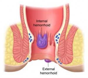
Hemorrhoids are swollen blood vessels of the rectum. The hemorrhoidal veins are located in the lowest area of the rectum and the anus. Sometimes they swell so that the vein walls become stretched, thin, and irritated by passing bowel movements. Hemorrhoids are classified into two general categories: internal and external.

Publish Post
Hemorrhoids


Internal hemorrhoids lie far enough inside the rectum that you can't see or feel them. They don't usually hurt because there are few pain-sensing nerves in the rectum. Bleeding may be the only sign that they are there. Sometimes internal hemorrhoids prolapse, or enlarge and protrude outside the anal sphincter. If so, you may be able to see or feel them as moist, pink pads of skin that are pinker than the surrounding area. Prolapsed hemorrhoids may hurt because the anus is dense with pain-sensing nerves. They usually recede into the rectum on their own; if they don't, they can be gently pushed back into place.
External hemorrhoids lie within the anus and are usually painful. If an external hemorrhoid prolapses to the outside (usually in the course of passing a stool), you can see and feel it. Blood clots sometimes form within prolapsed external hemorrhoids, causing an extremely painful condition called a thrombosis. If an external hemorrhoid becomes thrombosed, it can look rather frightening, turning purple or blue, and could possibly bleed. Despite their appearance, thrombosed hemorrhoids are usually not serious and will resolve themselves in about a week. If the pain is unbearable, your doctor can remove the thrombosed hemorrhoid, which stops the pain, during an office visit.
Anal bleeding and pain of any sort is alarming and should be evaluated; it can indicate a life-threatening condition, such as colorectal cancer. Hemorrhoids are the No. 1 cause of anal bleeding and are rarely dangerous, but a definite diagnosis from your physician is a must.

What Causes Hemorrhoids?

About half of the people in the U.S. will suffer from hemorrhoids at some point in life; for most, this will happen between ages 20 and 50. Researchers are not certain what causes hemorrhoids. "Weak" veins -- leading to hemorrhoids and other varicose veins -- may be inherited.
It's likely that extreme abdominal pressure causes the veins to swell and become susceptible to irritation. The pressure can be caused by obesity, pregnancy, standing or sitting for long periods, straining on the toilet, coughing, sneezing, vomiting, and holding your breath while straining to do physical labor.

Diet has a pivotal role in causing -- and preventing -- hemorrhoids. People who consistently eat a high-fiber diet are less likely to get hemorrhoids, but those who prefer a diet high in processed foods can expect them. A low-fiber diet or inadequate fluid intake can cause constipation, which can contribute to hemorrhoids in two ways: It promotes straining on the toilet and it also aggravates the hemorrhoids by producing hard stools that further irritate the swollen veins.
Internal External Hemorrhoids Image


Hemorrhoid Symptoms:
Hemroids (also called hemorrhoids), refer to the medical condition where blood vessels in the hemorrhoidal tissues of the rectal area become swollen, sensitive, inflamed or even bleeding. No one description of hemorrhoids symptoms fits all cases, since each case may vary from one patient to another. Usually, a variety of conditions present themselves on a case-by-case basis. Also, hemorrhoid symptoms depend on the type of hemroids you have, and the degree of their severity.
Two main categories of hemorrhoids are internal hemroids and external hemroids. There may be other conditions that present themselves, such as anal fissures, abscesses, itching, sensitivity, burning, and irritation of the entire anal or rectal areas. These are the commonly reported symptoms of hemroids.

How to Choose the Best Hemorrhoid Treatment

Hemorrhoid Treatment Reviews:
The very best hemroid treatment options require a robust level of screening to include not only the actual efficacy of the treatment, but also the safety of the product, the side effects if any, cost, handling, and any other brand-related issue like trust and value that should be considered. Millions of individuals are searching for the right kind of hemroid products—out of so many that are out there, which one is right for you? In each of our reviews, we’ve enlisted the following criteria to ensure you get a hemroid product that is meets your expectations for effectiveness, quality, value, and hemorrhoid relief. These criteria include:
Hemorrhoid Safety Image 


Safety
You’ll want to be ensured of the product’s safety, so it’s one of our most important levels of criteria in getting a product that works, but is also safe. Hemorrhoid treatment options that are not fully tested do not meet our safety standards. Only the hemorrhoid products that come packed with lab testing are included in our product reviews.
Effectiveness
Of course, one of the most significant reasons to use a hemorrhoid treatment is to resolve your issue and get instant relief. We’ve made sure to test and recommend only the products that actually worked and provided relief almost immediately. After all, no one wants to wait around for a hemroid treatment to actually work.
Cost
We make sure that our recommended products don’t cost you an arm and a leg! All of our recommended products offer a budget-friendly price that is both manageable and easy to accommodate. So, you’ll never find outrageously priced hemorrhoid treatment products that are simply unaffordable.
Customer Service
When you want answers, you’ll get them. With award-winning customer service teams, all of our recommended products are backed by a great team of customer service agents that are ready to assist you when you need it. All of our hemroid products offer the very best customer service.
Shipping Time
Hemorrhoids happen almost instantly, so you’ll need the shipping options that’ll actually work for you. No waiting around for weeks! All of our product reviews ensure that shipping options are there to get your hemorrhoid treatment to you in no time.

Hemorrhoid Relief:

How to Get Alternative Hemorrhoid Relief:
Hemorrhoids Cycle Photo
Hemorrhoids (known as hemroids) are a medical condition found on the anal and rectal areas. Luckily, hemroids are not serious, and they can be treated easily using a variety of treatments that offer hemorrhoid relief. Options range from natural supplements, hemorrhoid creams, surgery, and lifestyle changes. While any of these options can provide hemorrhoid relief, there are many alternative hemorrhoids treatments that can be just as effective—if not more effective—than other hemorrhoid cures.

Alternative Hemorrhoid Relief Products—How to Find the Best Treatment
If you’re looking for the best alternative hemorrhoid treatments, including at-home remedies, you should look for a product that not only deals with hemorrhoid symptoms but simultaneously provides hemorrhoid relief.
Any method that only treats hemorrhoid symptoms, however, won’t provide the comprehensive, permanent result you are looking for. You should find a two-part alternative treatment that can provide both instant and long-term benefits. Let’s discuss the lifestyle options first.
Fiber to the Diet
  • Aids in passing stool
  • Softens stool
  • Adds bulk
  • Reduces straining
One of the most popular alternative treatments for providing hemorrhoid relief includes adding fiber to your diet. You can do so naturally, by eating more of fruits, vegetables, whole grain pasta, whole grain cereals, whole grain breads, and any kind of bean or legume. This is the healthiest way to add fiber simply because it’s healthy as well. You can also supplement your diet with fiber supplements that use Psyllium husk to help add bulk to your stool.
Did you know that avoiding alcohol, cola drinks, and all types of caffeine helps in reducing hemorrhoids? Alternative hemorrhoid treatments that pertain to lifestyle include reducing your overall consumption of sugar, cheese products, spicy foods, greasy or high-fight foods, red meats, fatty nuts and even whole dairy products. You can eat these foods in moderation, but they key is to start adding whole, nutritious foods to your diet.
Other Alternative Hemorrhoid Relief Treatments

  •   Exercise


    • Exercise will provide the long-term hemorrhoid relief you are looking for. And, you’ll even look better as a result. What’s stopping you? Start exercising now. Even simple walking will do the trick.
      Hemorrhoids Exercise Image
      Try including exercise as part of your regular lifestyle routine. This will greatly aid in providing hemorrhoid relief and combating symptoms associated with painful, annoying hemroids. Try adding in a 20 – 30 minute walk, 3-5 times a day to your morning routine. Even light exercise can aid in improving bowel movements overall.
      You should try varied combinations of alternative treatments to find out what makes you feel better and what will eventually provide you with the greatest hemorrhoid relief. Not everyone reacts to foods, fiber, and exercise in the same way.
      • Natural-fiber underwear
      Natural fibers help to increase oxygen flow to the sensitive anal areas. You need to have air flow to aid in the proper balance of moisture contained in the anus region. Using natural fibers is a great way to encourage moisture control and to aid in drying the area out.
      Avoid nylons or synthetic materials that can trap moisture and make you feel even more uncomfortable. For women, using thongs is fine, but go for cotton panties occasionally if you suffer from regular hemroid symptoms. Making this simple switch in your daily lifestyle will aid your overall hemorrhoid relief strategy.

      Hair Loss

      Hair Loss:
      Now a days hair loss is a common problem for most of the people specially younger generation.Most of the people complain of lossing a lot of hair everyday.Hair loss is mainly due to genetic and environmental factors.

      Types of Hair Loss: 

      1. Alopecia areata.
      2. Androgenic Alopecia.
      3. Telogen Effluvium
      Alopecia areata

      Definition:Alopecia 
Areata Alopecia areata is the name for a condition in which round patches of hair loss appear suddenly. The hair-growing tissue is attacked by the patient's own immune cells for unknown reasons. There are three stages: first, there is sudden hair loss, then the patches of hair loss enlarge, and last, new hair grows back. This process takes months, sometimes more than a year, but rarely does the hair never grow back.


      Causes:
      • We do not know why the immune cells attack the hair-growing tissue.
      • Alopecia areata is not contagious, not caused by foods, and is not the result of nervousness.
      • Alopecia areata sometimes runs in families.
        Treatment:
      • Many people have achieved success with treatments such as Provillus, an all-natural alopecia solution for men and women.
      • Cortisone injections can also stimulate hair regrowth. Twenty to thirty injections per patch are required once a month. The injections are uncomfortable. Cortisone injections only work in the areas that have been injected; the injections do not prevent new areas of hair loss. It generally takes one to two months after the injection before hair growth is visible.
      • Some patients do not respond to any treatment. We cannot predict which patients will respond to treatment.
      • Cortisone creams are also sometimes beneficial in the treatment of alopecia areata.
      Androgenic Alopecia

      Definition:
      Androgenic alopecia is the name for male and female pattern baldness. In androgenic alopecia the hair loss occurs slowly over years. It can start anytime after age twenty. There is usually a family history of hair loss. In women, the hair slowly becomes thin throughout the scalp and bald spots usually do not occur. Men first develop hair loss at the temples, followed by an enlarging bald spot on top of the head.

      Causes:
      • Androgenic alopecia is thought to be due to the hair growing tissue's sensitivity to hormones; this sensitivity is due to genetic factors.
      • Not contagious, not caused by foods, not the result of nervousness.
      • Androgenic alopecia often runs in families.
      Treatment:
      • Rogaine (minoxidil) is a medicine used to treat high blood pressure which when applied to the skin causes hair growth in some individuals. One out of ten people who use Rogaine will grow hair. Rogaine will slow the rate of hair loss in five out of ten patients who use it. The other four out of ten are wasting their money.
      • Many people have achieved success with natural alternatives such as Provillus and other hair loss treatments.
      • Hair transplants are another option for treating androgenic alopecia. Plugs of hair are removed surgically from areas with plenty of hair and implanted in the areas with little hair.
      • Some people can benefit from scalp reduction. This is a surgical procedure in which the bald area is cut out.
      • Propecia is a new medication taken by mouth which treats androgenic alopecia.

      Telogen Effluvium

      Definition:
      Telogen effluvium is an abnormal loss of hair due to alteration of the normal hair cycle. Hair growth occurs in a cycle which is divided into three phases: a growth phase, a regression phase and a resting phase. Different hairs are in different stages of the cycle at any one time. Normally, most of the hairs are in the growth stage and only one hundred hairs per day fall from the scalp. When telogen effluvium occurs, a greater proportion of the hairs enter the resting phase of the cycle and hair shedding is greater than normal.

      Causes:
      • During pregnancy, more hair follicles are maintained in the growth phase. After pregnancy, a greater proportion of these hairs go into the resting phase, causing a temporary self-correcting increased shedding of the hair.
      • The same phenomenon can be seen after stopping birth control pills.
      • The flu or stress can also cause telogen effluvium.
      Treatment:
      • There is no treatment for telogen effluvium. The problem will correct itself. All the lost hair will eventually be replaced.